There’s a phenomenon that some people notice after major weight loss, but almost never have a scientific reasoning for it. Someone loses a large amount of body fat — 50 pounds, 100 pounds, sometimes more — and over time they notice something unexpected: Their complexion looks lighter. Not pale exactly, and not sickly, but fairer. Sometimes the change is subtle. Sometimes friends or family notice it before they do.
The phenomenon is largely anecdotal, among melanated people especially, stories about skin tone shifting after significant fat loss are surprisingly common. Usually, the explanation gets dismissed as lighting, reduced sun exposure, anemia, or simple perception. However, over the past two decades, researchers studying obesity, adipose tissue biology, inflammation, and pigmentation have uncovered several biological connections that make this observation far less mysterious than it first appears. What began as research into obesity-related inflammation has evolved into a broader understanding of how adipose tissue communicates with the immune system, oxidative stress pathways, and even melanogenesis — the biological process responsible for melanin production.
Read more below to find out why losing weight may cause your skin to lighten and how scientist are exploring the use of extracellular vesicles from fat cells to develop cosmetic therapeutics to combat hyperpigmentation and skin aging.
The Link Between Fat Tissue and Pigmentation
Adipose tissue is not “inactive” fat. For most of medical history, adipose tissue (fat), was regarded simply as excess energy storage, resulting from over consumption of food (dietary fats, sugars, and excess calories). Overtime, researchers began studying obesity at the molecular level. Now, adipose tissue is recognized as an active endocrine and immunological organ.
In the endocrine and immune systems, fat tissue produces hormones, cytokines, inflammatory mediators, and signaling molecules capable of influencing nearly every major body system. The more fat you gain, the larger adipose tissue becomes, particularly visceral fat — the fat surrounding organs. Excessive adipose tissue becomes more metabolically active and more inflammatory, which explains why obesity is strongly associated with chronic low-grade systemic inflammation, oxidative stress, insulin resistance, altered hormone signaling, and immune dysfunction.
In particular, inflammation and oxidative stress directly influence pigmentation.
Melanin is not just a pigment. For a long time, melanin was thought to exist primarily in specialized pigment-producing cells called melanocytes. These cells are found mainly in the skin, hair follicles, and eyes, and melanin is best known for determining skin and hair color. However, biologically, melanin also possesses potent antioxidant and anti-inflammatory properties. Interestingly, these properties play an important role in the relationship between fat and melanin production.
In 2010, researchers proposed that adipose tissue itself may participate in melanogenesis. In their review Melanin and melanogenesis in adipose tissue: possible mechanisms for abating oxidative stress and inflammation?, Page, Chandhoke, and Baranova examined evidence showing that adipose tissue expresses several genes related to melanogenesis and may produce melanin in response to metabolic stress. Researchers found that:
- Obesity is strongly associated with chronic low-grade inflammation
- Obesity is associated with increased oxidative stress
- Visceral adipose tissue from obese individuals contained more melanin compared to lean individuals
These finding led the researchers to hypothesize that melanin may function as a protective antioxidant defense mechanism inside adipose tissue . In other words, the body may increase melanogenesis in fat tissue as a compensatory response to chronic metabolic stress.
This theory may help explain why some individuals with obesity develop increased pigmentation or darker complexions over time — particularly when obesity is accompanied by systemic inflammation and insulin resistance.
Leptin, the Brain, and Melanin
Melanin is also expressed in certain regions of the central nervous system (brain). The Hypothalamus produces a neuropeptide known as melanin-concentrating hormone (MCH) which is important for regulating feeding behavior and energy balance. Interestingly, researchers discovered that adipocytes (fat cells that make up adipose tissue) also possess receptors for MCH.
In adipose tissue, MCH plays a role in the production and function of Leptin. One of the most important hormones produced by adipose tissue, leptin is a hormone that regulates appetite and satiety. This action is mediated by MCH through the hypothalamus.
Leptin also has major immune and inflammatory functions. Increased body fat raises leptin levels, and high leptin level promote inflammatory cytokine production. In turn, inflammatory cytokines can further increase leptin production. This creates a self-perpetuating inflammatory feedback loop frequently seen in obesity.
These findings suggest that pathways involved in pigmentation, appetite regulation, neuroendocrine signaling, and adipose biology may all intersect.The relationship between adipose tissue and melanin associated pigmentation therefore appears to involve, oxidative stress, inflammation, endocrine activity, and melanogenic gene expression.
Inflammation, Insulin Resistance, and Hyperpigmentation
Excess fat creates a chemical microenvironment that encourages pigmentation. As we saw, fat tissue produces inflammatory cytokines. These cytokines can alter insulin signaling leading to insulin resistance and increase metabolic stress. Metabolic stress increases oxidative stress and more inflammation, and all of these systems intersect with melanogenesis. Lets break down how:
One of the clearest clinical examples connecting obesity and pigmentation is a condition called acanthosis nigricans.
Acanthosis nigricans is a hyperpigmentation disorder that commonly appears in individuals with obesity, insulin resistance, Type 2 diabetes, and metabolic syndrome. People with the condition develop thick, darkened, velvety patches of skin – usually localized around the neck, armpits, groin, and body folds, unlike generalized skin darkening. However, Acanthosis nigrican demonstrates an important principle: Metabolic dysfunction and chronic inflammation can visibly influence pigment production and skin cell growth.
Hyperinsulinemia associated with insulin resistance stimulates pathways that increase keratinocyte proliferation and melanocyte activity. Simultaneously, oxidative stress and inflammatory cytokines can further amplify melanogenesis. As obesity is often responsible for insulin resistance and Type 2 Diabetes, excess fat may thereby create a biological environment that promotes increased pigmentation signaling throughout the body.
Why Weight Loss May Ligthen Complexion
Although the scientific literature has not definitively proven that fat loss causes lighter skin tone, several mechanisms make the observation biologically plausible.
If obesity increases systemic inflammation, oxidative stress, insulin resistance, leptin, and potentially compensatory melanin production, then major fat loss may reduce those same signals. Weight loss alters systemic inflammation, oxidative stress, and hormonal and cytokine signaling.
As inflammation and oxidative stress are known stimulators of melanogenesis, reducing these signals may lowers the body’s overall demand for melanin production. Also, since obesity increases reactive oxygen species (ROS) production, and melanin acts as a scavenger of ROS, reducing adipose tissue could theoretically reduce oxidative stress and consequentially, compensatory melanogenesis. Additionally, as leptin, insulin, and adipose cytokine secretion by fat issue intersects with inflammatory and melanogenic pathways, changes in body fat may indirectly influence pigmentation.
In simpler terms:
Inflammatory burden goes down, insulin sensitivity improves, oxidative stress decreases, circulating leptin is lowered, and there is overall less chronic metabolic stress. Less metabolic stress may mean less need for melanin-related protective activity.
If melanogenesis is functioning partly as a protective response to oxidative and inflammatory stress, then reducing the underlying stress environment could theoretically reduce melanogenic activity as well. This may be enough to produce visible changes in complexion for some people following dramatic weight loss. Although this change may not occur overnight or as dramatically in every individual, it is possible.
A New Frontier: Adipose Stem Cellular Vesicles
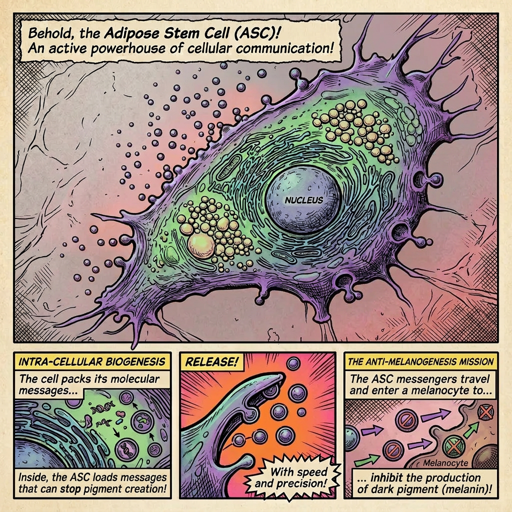
While dysfunctional adipose tissue may contribute to inflammatory stress, scientists are now discovering that biological nanoparticles released from adipose stem cells may possess the opposite effect.
These particles are called extracellular vesicles, lipid-bound particles releases by cells that contain microRNA’s, proteins, and lipids. EVs act as communication systems between tissues and organs and help regulate cellular homeostasis. Interestingly, small EVs (~200 nm in diameter) often perform functions that differ from — or even oppose — those of their parent cells.
In 2025, emerging research on adipose-derived small extracellular vesicles investigated their use in cosmetic therapeutics. Researched assessed adipose stem cell small EVs (ASC-sEVs) as delivery systems for natural antioxidant compounds. Remarkably, researchers discovered that ASC-sEVs demonstrated anti-melanogenic effects in melanocyte cells.
By themselves, the vesicles reduced melanin production, but when loaded with antioxidant compounds like resveratrol and arbutin, anti-melanogenic effects became significantly stronger. Likely, due to the vesicles’ ability to improve skin penetration of the antioxidant compounds. Additionally, the vesicles enhanced compound stability, bioavailability, and intracellular delivery while requiring lower therapeutic concentrations than the antioxidants alone. Importantly, the treatments were not cytotoxic.
This introduces a fascinating paradox: while obesity-associated adipose tissue may contribute to inflammatory melanogenic signaling, extracellular vesicles derived from adipose stem cells may simultaneously possess regulatory and anti-pigmentary functions capable of restoring balance.
Timeline of the Science Connecting Adipose Tissue, Inflammation, and Melanin
Pre-2000s 2000 Mid-2000s 2008–2011 2010s 2021 2025 Present ●──────────────────●────────────────────●────────────────────●────────────────────●────────────────────●────────────────────●────────────────────● Passive fat MCH receptors Obesity viewed Melanin found Insulin resistance Obesity reframed ASC-sEVs shown Integrated model storage model discovered in as inflammatory in adipose tissue linked to as neuroimmune to reduce emerging: adipocytes condition + melanogenesis hyperpigmentation endocrine disorder melanogenesis fat, inflammation, genes identified + enhance oxidative stress, antioxidant delivery pigmentation linked together ↓ ↓ ↓ ↓ ↓ ↓ ↓ Fat tissue Cytokines, ROS, Melanin proposed Acanthosis Leptin linked Adipose stem-cell Weight loss may linked to leptin, and as antioxidant nigricans shows to inflammation, EVs explored as reduce systemic neuroendocrine insulin resistance defense mechanism metabolism can immunity, and regenerative melanogenic signaling become central against oxidative alter pigmentation metabolic therapeutics signaling through stress homeostasis lower inflammation
Conclusion
The relationship between body fat and skin pigmentation is far more biologically complex than previously understood.
Emerging evidence suggests that obesity-associated inflammation, oxidative stress, insulin resistance, and endocrine signaling may all influence melanogenesis both locally and systemically.
This may help explain why some individuals notice skin lightening after major fat loss: reduced adipose burden may decrease inflammatory and oxidative signals that promote compensatory pigmentation activity.
At the same time, modern research is revealing a fascinating duality within adipose biology itself. While dysfunctional adipose tissue can contribute to chronic inflammation, adipose-derived extracellular vesicles may possess powerful regenerative and anti-melanogenic properties capable of restoring cellular balance.
What once seemed like a cosmetic anecdote may ultimately become part of a much broader scientific story connecting metabolism, pigmentation, inflammation, and regenerative medicine.
References
Bradley, R. L., Kokkotou, E. G., Maratos-Flier, E., & Cheatham, B. (2000). Melanin-concentrating hormone regulates leptin synthesis and secretion in rat adipocytes. Diabetes, 49(7), 1073–1077. https://doi.org/10.2337/diabetes.49.7.1073
Page, S., Chandhoke, V., & Baranova, A. (2011). Melanin and melanogenesis in adipose tissue: Possible mechanisms for abating oxidative stress and inflammation? Obesity Reviews, 12(5), e21–e31. https://doi.org/10.1111/j.1467-789X.2010.00773.x
Procaccini, C., La Rocca, C., Carbone, F., & Matarese, G. (2021). Leptin and obesity: Role and clinical implication. Frontiers in Endocrinology, 12, 585887. https://doi.org/10.3389/fendo.2021.585887
Vo, N., Vu, D. M., Tran, N. H. B., Nguyen, D. D. N., Phung, P. M., Nguyen, H. N., & Tu, L. N. (2025). Synergistic anti-aging effects of adipose-derived stem cell extracellular vesicles loaded with natural compounds. Journal of Cosmetic Dermatology, 24(2), e70021. https://doi.org/10.1111/jocd.70021


 There is a common misconception that the popular energy drink RedBull contains urine or sperm from bulls, because it contains taurine. The myth stems from the fact that taurine was originally isolated from Ox bile (bulls and oxen are male cows) in the 1800’s. While initially identified in the bile (produced by the liver) of male cows, taurine is widely found in almost every organ in mammals. Taurine is chemically synthesized and used as an ingredient in infant formula, energy drinks, nutritional supplements, and pet food. Most recently it has been identified for its therapeutic value in the treatment of congestive heart failure and other cardiovascular diseases. It is currently being studied for its further use in the treatment of other diseases, in particular those relating to the nervous system, muscles, and mitochondrial disorders.